Two Cases of Kawasaki Disease Presenting with Meningitis
Article information

Kawasaki disease is a pediatric systemic inflammatory vasculitis first described in Japan by Dr. Tomisaku Kawasaki in 1967 [1]. The etiology of Kawasaki disease remains uncertain, with hypotheses ranging from genetic predisposition to infectious agents [2]. A diagnosis of complete Kawasaki disease is established when a patient has a fever for 5 or more days, along with at least four of the following five symptoms: bilateral conjunctival injection, changes in the oral mucosa, cervical lymphadenopathy, changes in the extremities, and polymorphous rash [3]. Incomplete Kawasaki disease is considered when a patient presents with a fever lasting 5 or more days but exhibits fewer than four of the clinical signs [4]. The highest incidence is observed in toddlers, with only rare cases in infants younger than 3 months of age. Neurological complications are recognized in 1% to 30% of Kawasaki disease cases [5]. These complications include irritability, aseptic meningitis, lethargy, transient hemiplegia, cerebral infarction, ataxia, seizures, and focal encephalopathy [6]. This report presents two unusual cases of Kawasaki disease in infants who were initially diagnosed and treated for meningitis but were later confirmed to have Kawasaki disease.
Case 1: A previously healthy 2-month-old boy presented with a 3-day history of fever. The parents did not report any additional symptoms. Physical examination revealed no noteworthy findings. Initial blood tests indicated a white blood cell count of 8.42×10⁹/L (with 52.4% neutrophils), a hemoglobin level of 9.6 g/dL, and a platelet count of 207×109/L. The inflammatory marker C-reactive protein (CRP) was elevated at 13.1 mg/dL (reference range, <0.5), while routine biochemistry, liver function, and renal function tests were unremarkable. Cerebrospinal fluid (CSF) analysis indicated a cell count of 120/mm3 (20% mononuclear cells and 80% polymorphonuclear cells), a protein level of 80.6 mg/dL, and a glucose level of 63 mg/dL (serum glucose, 129). CSF cultures and polymerase chain reaction tests—including those for enterovirus, herpes simplex virus types 1 and 2, varicella-zoster virus, and human herpesvirus 6—were negative. Brain magnetic resonance imaging revealed nonspecific findings. The patient was initially treated with intravenous antibiotics (specifically, ampicillin, and cefotaxime) under the suspicion of bacterial meningitis. However, blood, CSF, and urine cultures yielded negative results, and the fever persisted for 7 days. On the 6th day of admission, follow-up testing revealed leukocytosis (23.43×10⁹/L), thrombocytosis (605×109/L), and elevated measurements for CRP level (7.01 mg/dL), erythrocyte sedimentation rate (ESR) (99 mm/hr; reference range, 0 to 20), and N-terminal pro B-type natriuretic peptide (NT-proBNP) level (3,489 pg/mL; reference range, 0 to 125) (Table 1). On the 5th day of hospitalization, the patient developed bilateral conjunctivitis, cracked and erythematous lips, and redness at the bacillus Calmette-Guérin (BCG) vaccination site and anus (Fig. 1). Echocardiography revealed no abnormalities. The patient was ultimately diagnosed with Kawasaki disease, as he met the criteria of persistent fever (lasting 7 days), bilateral conjunctival injection, changes in the oral mucosa and extremities, and polymorphous rash. Treatment with intravenous immunoglobulin (IVIG) (2 g/kg/dose) and high-dose acetylsalicylic acid (50 mg/kg/dose for 3 days) was initiated. Following IVIG administration, the fever resolved, and subsequent tests showed decreased levels of CRP (1.23 mg/dL) and NT-proBNP (282.5 pg/mL) (Table 1). After 10 days of hospitalization, the patient was discharged on a low-dose acetylsalicylic acid regimen (5 mg/kg/dose) with a cardiology follow-up scheduled. At the 3-month follow-up, no abnormalities or complications were noted.

Laboratory findings for the two cases
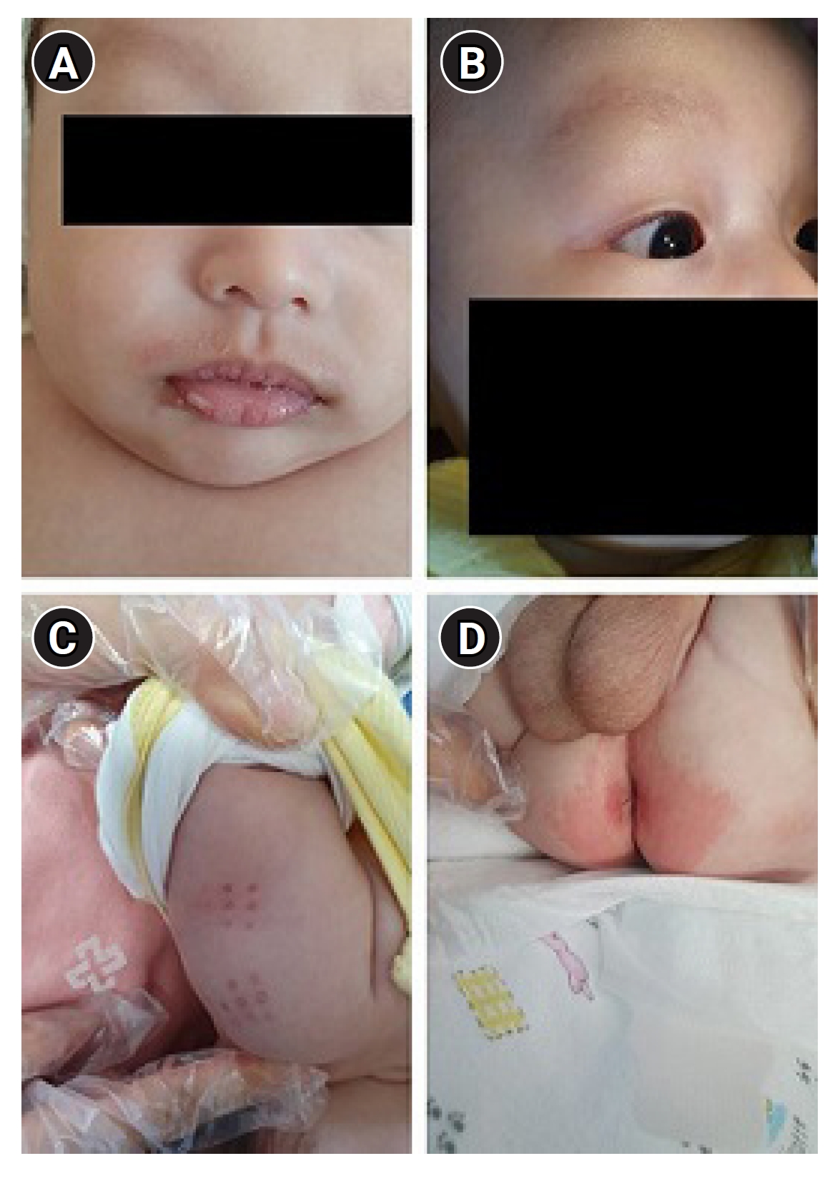
Case 1 showing Kawasaki features such as cracked and erythematous lip (A), bilateral conjunctivitis (B), bacillus Calmette-Guérin (BCG) site redness (C), and anus redness (D).
Case 2: A 3-month-old boy presented with a 2-day history of fever. No specific accompanying symptoms or signs were reported, and the physical examination revealed no noteworthy findings. Laboratory tests indicated a white blood cell count of 11.15×10⁹/L (with 44.4% neutrophils), a hemoglobin level of 10.0 g/dL, and a platelet count of 329×109/L. The patient’s CRP level was elevated at 3.26 mg/dL, but routine biochemistry, liver, and renal function tests yielded normal results. CSF analysis revealed a cell count of 60/mm3 (70% mononuclear and 30% polymorphonuclear cells), a protein level of 41.8 mg/dL, and a glucose level of 75 mg/dL (serum glucose, 128 mg/dL) (Table 1). Considering the possibility of bacterial or viral meningitis, the patient was treated with intravenous antibiotics (vancomycin and cefotaxime) and acyclovir. The fever briefly subsided on the second day of admission but recurred on the 6th day. The patient exhibited classic signs of Kawasaki disease, including persistent fever for 5 days, bilateral conjunctivitis, red lips, redness at the BCG vaccination site, rash, and swelling of both hands. Echocardiography showed no abnormalities, including of the coronary artery. On the 6th day of admission, follow-up laboratory tests indicated leukocytosis (12.54×10⁹/L), along with elevated levels of CRP (9.23 mg/dL) and NT-proBNP (1441.6 pg/mL) (Table 1). Kawasaki disease was suspected, and the fever resolved after treatment with IVIG and high-dose acetylsalicylic acid. On the 9th day of admission, blood testing revealed a white blood cell count of 6.40×10⁹/L, a normalized CRP level of 0.08 mg/dL, and a markedly reduced NT-proBNP level (90.5 pg/mL). The patient was discharged on a low-dose acetylsalicylic acid regimen. Follow-up echocardiography at 2 and 9 months after discharge produced normal results, with no coronary artery or neurological complications.
Written informed consent was waived due to the retrospective nature of the study. The research was approved by the Institutional Review Board of Busan Paik Hospital (Busan, Korea; approval number: 2022-10-062).
Kawasaki disease is diagnosed based on clinical manifestations. A less prominent clinical picture can easily lead to a missed diagnosis. Incomplete Kawasaki disease, most commonly observed in infants, poses a substantial risk of coronary artery abnormalities. These infants may present with prolonged fever as the only clinical finding, or they may exhibit subtle clinical signs in addition to fever [7]. In our cases, the patients were less than 3 months old, and fever was initially the only symptom observed. Rather than appearing simultaneously, clinical features of this condition may evolve sequentially over time, as seen in our cases. Kawasaki disease can manifest in the central nervous system, with symptoms including seizures, facial paralysis, hemiplegia, myositis, and aseptic meningitis [8]. Aseptic meningitis is an uncommon feature of Kawasaki disease, so its presence in patients with Kawasaki disease can complicate accurate and timely diagnosis [7]. Therefore, in cases of aseptic meningitis that do not respond to antibiotics or antiviral treatment, as with our patients, Kawasaki disease should be suspected. While no specific diagnostic test is available for this condition, certain laboratory findings can support the diagnosis of incomplete Kawasaki disease. During the acute phase, observations may include leukocytosis, anemia, hypoalbuminemia, elevated ESR, and elevated levels of CRP, serum transaminases or gamma-glutamyl transpeptidase, and NT-proBNP. Thrombocytosis occurs during the subacute phase; however, these findings are less useful for diagnostic purposes [4].
We present two cases of Kawasaki disease presenting with meningitis. The initial symptom in both cases was an isolated fever, with subsequent symptoms and laboratory findings that confirmed Kawasaki disease associated with aseptic meningitis. The diagnosis was complicated by the patients’ young age, the lack of symptoms other than fever, and the gradual appearance of features characteristic of Kawasaki disease. If not diagnosed promptly, Kawasaki disease can result in numerous complications. Thus, this condition should be considered in young infants with meningitis that does not respond to treatment.
Notes
No potential conflict of interest relevant to this article was reported.
Author contribution
Conceptualization: SK and BLL. Data curation: SK and BLL. Methodology: SK and BLL. Project administration: SK, JEL, DER, and BLL. Visualization: SK and BLL. Writing-original draft: SK and BLL. Writing-review & editing: SK, JEL, DER, and BLL.