 |
 |
- Search
| Ann Child Neurol > Volume 29(4); 2021 > Article |
|
Hypoglossal nerve (cranial nerve XII) palsy (HNP) is a neurological disorder that causes tongue deviation, atrophy, and muscle fiber contraction on the affected side. Tumors are the most common cause of HNP, followed by trauma, vascular abnormalities, autoimmune diseases, and rarely, viral or bacterial infection [1,2]. Isolated HNP without other neurological symptoms is rare. It is often accompanied by other cranial nerve disorders; intramedullary lesions typically involve the adjacent nuclei or tracts, and peripheral lesions generally affect other lower cranial nerves [1]. Furthermore, isolated HNP following influenza virus infection has not been reported previously. This report describes a case of isolated HNP associated with influenza B virus infection in a child.
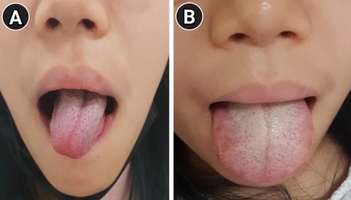
A 12-year-old girl visited our hospital with tongue deviation to the right on protrusion and involuntary tongue movement. She had a history of crustacean allergy. Except for stomatitis 1 month prior, the patient had no history of recent trauma or surgery, as well as no significant family history. She had developed a fever 3 days before the onset of abnormal tongue movement. On the first day of fever onset, she was diagnosed with influenza B virus infection through a rapid influenza antigen test with nasopharyngeal specimens and started oseltamivir therapy (60 mg twice a day; the recommended dosage for a weight of 38 kg). Her fever subsided on the second day after oseltamivir administration. She complained of pain in both calves on the third day of treatment with oseltamivir. On day 4, a posterior neck muscle was pulled during treatment with oseltamivir, and she displayed involuntary tongue protrusion and deviation to the right on protrusion. Atrophy and fasciculation of the tongue were not observed (Fig. 1). No involvement of other cranial nerves was detected.
Her body temperature was 36.9┬░C, her heart rate was 72 bpm, and her blood pressure was 110/60 mm Hg. Laboratory tests revealed leukopenia, with a white blood cell count of 2,230/mm3, and the other laboratory test results were as follows: hemoglobin, 13.2 g/dL; platelets, 113,000/mm3; aspartate aminotransferase, 132.9 IU/L; alanine aminotransferase, 32.4 IU/L; C-reactive protein, 0.13 mg/L; creatine kinase, 5,148 U/L; myoglobin, 414.1 ng/mL; and lactate dehydrogenase, 734 U/L. Serum anti-cytomegalovirus immunoglobulin (Ig) M and IgG, serum Epstein-Barr virus IgM and IgG, and serum Mycoplasma IgM and IgG were all negative. The serum herpes simplex virus IgM titer was 1:4; however, herpes simplex virus was not detected in a polymerase chain reaction test. She also tested negative for anti-nuclear antibodies. Brain magnetic resonance imaging (MRI) involving thin sections of the skull base, including the medulla oblongata (where the hypoglossal nuclei are located), and magnetic resonance angiography (MRA) showed no pathological findings [3].
Thus, we diagnosed isolated HNP related to influenza B virus infection through exclusion [4]. She did not receive any specific therapy aside from oseltamivir for the influenza B infection. The tongue deviation on protrusion and involuntary tongue movement fully resolved the day after symptom onset.
Cranial nerve palsy after influenza B infection is rare [5], but oculomotor nerve palsy and trochlear nerve palsy after influenza B infection have been reported [6,7]. There was also a report of isolated HNP after influenza vaccination [4]. Several reports have described HNP caused by other viral or bacterial infections without broad structural abnormalities [2,4]. These cases were related to infections such as enterovirus, adenovirus, Epstein-Barr virus, herpes simplex virus, and Streptococcus spp. [3,8,9]. However, HNP related to influenza virus infection has not been reported previously in children or adults. To our knowledge, this is the first report of isolated HNP related to influenza B virus infection.
Infectious causes of cranial nerve palsy are postulated to involve either direct infection of the nerve or immune processes, but cranial nerve palsy can also occur due to nerve compression by reactive lymph nodes [3,9]. In the current case, there was no evidence of lymphadenopathy or specific structural abnormalities in hypoglossal nerve pathways on brain MRI and MRA. Therefore, it is speculated that hypoglossal nerve palsy was caused by direct infection of the nerve with influenza B virus or by immune processes.
We did not administer any specific treatment other than oseltamivir. When HNP developed, the patient had received treatment with oseltamivir for 4 days and continued the treatment. The involuntary movement and deviation of the tongue fully resolved the day after symptom onset. The disease course was thus shorter than in other cases [1-3,9].
The symptoms of HNP occurred during treatment with oseltamivir. However, cranial nerve palsy as a side effect of oseltamivir has not yet been reported. Further, the patient continued oseltamivir treatment after HNP developed, and the symptoms resolved during the course of medication. Therefore, we excluded the possibility that HNP was a side effect of oseltamivir.
HNP without structural abnormalities has a good prognosis [1,4]. Some patients receive oral prednisolone; however, most cases resolve without specific treatment [3]. Since post-infectious HNP is very rare, there is no established treatment. However, in the absence of other serious causes, observation of symptoms without further treatment seems to be sufficient.
In conclusion, when an apparent cause of HNP is not identified, a test for the detection of influenza virus should be conducted.
This study was approved by the Institutional Review Board of Chosun University Hospital (CHOSUN 2021-05-004) (IRB review exemption). Informed consent was waived by the board.
Notes
Author contribution
Conceptualization: SEY. Data curation: IBP and SEY. Formal analysis: IBP and SEY. Methodology: IBP and SEY. Project administration: SEY. Visualization: IBP. Writing-original draft: IBP and SEY. Writing-review & editing: SEY.
References
1. Combarros O, Alvarez de Arcaya A, Berciano J. Isolated unilateral hypoglossal nerve palsy: nine cases. J Neurol 1998;245:98-100.


3. Hadjikoutis S, Jayawant S, Stoodley N. Isolated hypoglossal nerve palsy in a 14-year-old girl. Eur J Paediatr Neurol 2002;6:225-8.


4. Shibata A, Kimura M, Ishibashi K, Umemura M. Idiopathic isolated unilateral hypoglossal nerve palsy: a report of 2 cases and review of the literature. J Oral Maxillofac Surg 2018;76:1454-9.


6. Kim HJ, Kim MS, Kim HS. Acute isolated oculomotor nerve palsy after influenza B virus infection in a 4-year-old girl. J Korean Child Neurol Soc 2016;24:178-82.

7. McCombe JA, Narayansingh MJ, Jhamandas JH. Cranial nerve palsies associated with influenza B. Can J Neurol Sci 2009;36:262-4.


- TOOLS
-
METRICS

-
- 0 Crossref
- Scopus
- 5,477 View
- 82 Download
- Related articles in Ann Child Neurol
-
Unilateral and Reversible Hypoglossal Nerve Palsy Due to Infectious Mononucleosis2022 January;30(1)
A Case of Brainstem Encephalitis Associated with Epstein-Barr Virus Infection.2011 December;19(3)


