Ann Child Neurol Search

CLOSE


Ann Child Neurol > Volume 29(2); 2021 > Article
Kim, Shim, Ko, Kim, Kim, Lim, Hwang, Choi, Kim, and Chae: Clinical and Genetic Spectrum of STXBP1 Encephalopathy in the Korean Pediatric Population

Abstract

Purpose

Syntaxin-binding protein 1 (STXBP1) mutations are known to result in various phenotypes including Ohtahara syndrome, West syndrome, and autism, collectively referred as STXBP1 encephalopathy. This study aimed to expand our understanding of the genotype-phenotype spectrum of STXBP1 encephalopathy in the Korean pediatric population.

Methods

Ten patients with STXBP1 mutations were enrolled for a retrospective chart review. The patients were investigated for developmental delay of unknown cause and epileptic encephalopathy at a single center.

Results

Ten different STXBP1 mutations were identified. Three mutations had not previously been reported (c.1212A>C, c.1497C>G, c1030-2A>G). Eight patients showed early-onset epileptic encephalopathy as the main feature, while the main feature was developmental delay and non-epileptic movements in two patients. The most commonly seen electroencephalographic change was focal/multifocal epileptiform discharges, which were observed in nine patients (90%). The classical burst-suppression pattern was observed in four patients, two of which evolved to show hypsarrthymia. All patients with seizures had drug-resistant epilepsy. The patients suffered from severe developmental delay regardless of seizure frequency. Six patients showed an associated movement disorder or behavioral disorder.

Conclusion

This study describes the STXBP1 encephalopathy patients in Korean pediatric population, further expanding knowledge of its phenotype spectrum.

Introduction

Developmental and epileptic encephalopathy is a concept to acknowledge that many genetic disorders show developmental impairment as a direct consequence of the genetic mutation, in addition to the detrimental effect of the frequent epileptic activity on brain development [1]. In the era of next generation sequencing, increasing monogenic causes for developmental and epileptic encephalopathy are being discovered, providing us with new insight to its underlying patho-genetic mechanisms.
Syntaxin-binding protein 1 (STXBP1) (also known as MUNC18-1) is a member of the membrane trafficking proteins predominantly expressed in the brain, which is important for docking and fusion of the synaptic vesicles [2]. There is approximately 200 cases reported in the literature until date, mostly being de novo heterozygous mutations [3]. STXBP1 mutation was first described in 2008, in patients with early infantile epileptic encephalopathy with suppression-burst (EIEE), or Ohtahara syndrome [4]. Subsequent studies showed that STXBP1 mutation is responsible for approximately 22% of cases with Ohtahara syndrome, 6% of non-syndromic early onset epileptic encephalopathy and 2% of West syndrome [4-7]. Due to the expanding phenotype STXBP1 encephalopathy was suggested to be more appropriate [8].
Recognition and classification of diverse phenotypes arising in STXBP1 mutation is crucial to guide future management options. Here, we report 10 patients with STXBP1 mutation from a single center and summarize the detailed clinical features, with the aim to expand its phenotypic spectrum in Korean pediatric population.

Materials and Methods

Patients with STXBP1 mutation were identified from a cohort of 198 pediatric patients with developmental and epileptic encephalopathy of unknown etiology. Patient cohort was selected from the Division of Pediatric Neurology of the Seoul National University Children’s Hospital from September 2012 to May 2019. All the patients had no obvious etiology based on clinical features, neuroimaging and metabolic screening. The first-tier test included chromosomal microarray or targeted multi-gene panel. Whole exome sequencing was conducted as a first-tier test as well as a second-tier test, in selected cases based on the decision of the child neurology expert consortium. Variants were evaluated and classified according to the guideline proposed by American College of Medical Genetics (ACMG) [9]. ClinVar database was searched for past variant reports [10]. Population frequency of variants were determined using 1000genome, ExaC, and gNomad database.
STXBP1 sequencing was performed on DNA from probands and family members using the Sanger method to identify parental origin whenever possible. Information obtained from medical records include age at seizure onset, symptoms at onset, duration from onset to diagnosis, previous diagnosis and treatment, associated psychiatric and behavior symptoms, response to antiepileptic drug (AED) treatment and developmental outcome. Each patient’s electroencephalography (EEG) was obtained and analyzed. Seizure types and epileptic syndromes were classified according to the 2017 International League Against Epilepsy guidelines [1].
The study protocol was approved by the Institutional Review Board of Seoul National University Hospital (IRB No.H-2001-134-1096), and the study was conducted in accordance with relevant guidelines and regulations. Written informed consent by the patients was waived due to the retrospective nature of our study.

Results

1. Genetic identification of STXBP1 mutations

Total of 10 different variants were identified in 10 patients (Table 1 and Fig. 1). Four variants were missense mutations, another four variants were nonsense mutations, and two variants were splicing mutations. Three novel variants (c.1212A>C, c.1497C>G, c.1030-2 A>G) and seven variants previously reported as pathogenic [3,8,11-15] were identified. All novel variants were not found in either 1000Genomes or ExAC control database. Total of eight patients’ parental DNA sample were available for segregation analysis. All eight patients tested harbored de novo mutation.

2. Clinical characteristic of STXBP1 mutations

The clinical characteristics of 10 patients with STXBP1 mutations are summarized in Table 1. Patient’s median age was 7 years old (range, 1 to 11). Head circumference was normal in all patients. Brain magnetic resonance imaging from all patients did not show remarkable abnormalities. Among 10 patients, nine patients had confirmed electroclinical seizures. The median age of seizure onset was 6 months old (range, 3 days to 7 years). Three patients showed neonatal onset seizures, presenting within 1 month of age and other five patients presented as early onset epilepsy, presenting with seizures before the age of 3 years. In remaining two patients seizure was not a main clinical feature; patient 6 was being followed up for global developmental delay and ataxia before his first seizure at age of 7 years. Patient 7 never had clinical seizures until the age of 8 years at last follow-up, but suffered from head dyskinesia, bruxism, and hand stereotypy.
All patients showed significant global developmental delay regardless of seizure frequency. All domains of development were delayed, but the language domain was always more severely affected compared to the motor domain. Only one patient was able to achieve any word output, whereas four patients achieved the milestone of walking. Three patients (patient 4, 6, 7) were already being followed for a global developmental disorder before seizure occurrence. Two patients (patient 6, 7), who’s seizure was not a dominant feature, showed clear developmental regression during follow-up. In patient 6, motor function deterioration was observed after seizure onset, which progressed to worsening ataxia and dysphagia. Patient 7 showed developmental regression being evident at the age of 2 years without any clinical seizures, which lead to initial misdiagnosis as Rett syndrome.
Six patients showed additional symptoms other than seizure. Behavior problem was seen in five patients showing autistic like features including hyperactivity/disruptive behavior (n=3), bruxism (n=2), hand stereotypy (n=2). Neurologic symptoms were seen in three patients, including tremor (n=2), ataxia (n=1), dystonia (n=1), hypotonia (n=1), and dyskinesia (n=1).
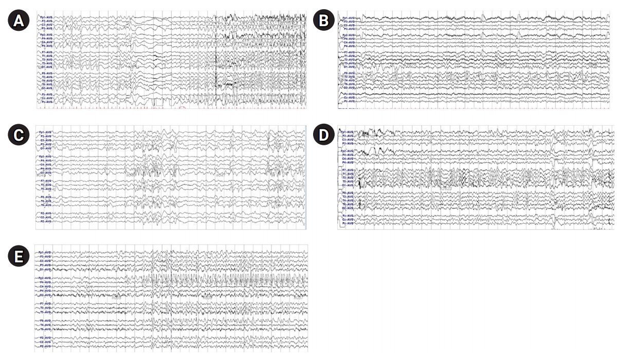
Four patients (patient 1, 3, 5, 8) were initially diagnosed as EIEE (or Ohtahara syndrome) with background burst-suppression pattern with multifocal spikes. Two patients (patient 3, 5) went on to develop hypsarrythmia with epileptic spasms and diagnosis was changed to West syndrome. A total of six patients (patient 1, 2, 4, 6, 9, 10) eventually evolved to unspecified epileptic encephalopathy with focal and multifocal spike discharges (Fig. 2). Patient 7 showed irregular high amplitude delta activities in the background activity. All patients except patient 7 (n=9) showed either focal or multifocal epileptiform discharges in EEG.
Tonic seizure was the most common type seen (n=8), but multiple types of seizure semiology are observed including epileptic spasms (n=4), myoclonic (n=2), and behavior arrest (n=2) seizure. Six patients overall showed more than one type of seizure semiology.
Regarding treatments, all patients with epilepsy were refractory to antiepileptic medications, requiring more than two types of AEDs, and none achieved seizure freedom through medication. Of note, patient 10 showed significance response to ketogenic diet, leading to cessation of all AEDs.

Discussion

Here we identified 10 Korean patients with STXBP1 mutation, which included three novel mutations. We described detailed phenotypes and genotypes of the patients with STXBP1 encephalopathy. Due to its rarity, the clinical spectrum of STXBP1 encephalopathy is not yet well known, but recent large cohort study suggests that intellectual disability and epilepsy is the two main major components of STXBP1 encephalopathy.
Epilepsy was observed in 95% of STXBP1 encephalopathy patients. Among the patients with available information, about half was taking more than three AEDs and one-third was suffering from frequent seizure. On the other hand approximately one-third achieved seizure freedom [3]. Consistent with the above observation, nine patients (90%) in the current cohort showed epilepsy and all patients required more than two AEDs.
In the same study, moderate to severe intellectual disability was observed in 88.4% of patient. Notably, developmental delay was present before seizure onset in 64.3%, and 7% of patients were observed to have just developmental delay without any seizure. Other previous studies also observed that some degree of developmental delay was often observed prior to any seizure onset, thus it is considered an independent domain from epilepsy [3,12]. Indeed, in our cohort all patients showed profound developmental delay or severe intellectual disability and 30% of patients showed developmental delay before seizure onset.
However, it is often difficult to assess whether seizure activity or epileptiform discharges have any effect on developmental process or whether developmental delay is totally separate phenotype in STXBP1 encephalopathy. It was suggested that developmental regression is rarely seen and does not seem to be related to seizure activity [3]. In the current cohort, patient 6 and 7 showed clear regression during follow-up. Interestingly, both patients were different from typical patients presenting with early onset epileptic encephalopathy, as seizure was not their main feature and both showed predominantly non-epileptic movement symptoms. Patient 7 followed Rett syndrome like features with regression starting at age of 3 with autistic features, and patient 6 showed many resemblances to the phenotype previously described as ataxia-tremor-retardation syndrome [16]. Patient 6 showed clear motor function regression after seizure onset with worsened ataxia and newly developed dysphagia. Above two patients support that seizure activity and intellectual development are indeed independent domains.
STXBP1 encephalopathy was known to be most commonly associate with burst suppression and hypsarrhythmia, as it was initially described mostly in Ohtahara syndrome [11,12,17]. However recent large cohort study suggest most commonly described changes are focal or multifocal activities, seen in 64% of the cases [3]. Our patients also showed that focal or multifocal epileptiform discharges were the most common finding (60%), and classical burst-suppression patterns were seen in only proportion of the patients (40%). However, patients who were followed up long term showed that patients with burst-suppression pattern often evolve to hypsarrhythmia or non-specific focal patterns. Thus it is possible some patients did have burst-suppression pattern at one point but was never recorded. The range of abnormal EEG findings was wide in the current cohort, and they showed little correlation with their developmental status or seizure activity, in accordance to previous reports [3,18].
The reason why the clinical presentation of patients with STXBP1 mutation varies so much is yet unknown. Previous evidence supported the hypothesis that haploinsufficiency is the main pathogenic mechanism underlying STXBP1 encephalopathy, which may explain for such phenotypic heterogeneity [19]. However, there is growing evidence that STXBP1 mutation have more than one mechanism of pathophysiology. Recently, a homozygous STXBP1 mutation was found to cause Lennox-Gastaut syndrome, showing that STXBP1 also have a dominant-negative effect [20]. Thus mechanism of STXBP1 encephalopathy still needs much further research in the future.
Another important aspect of STXBP1 mutation to consider is mosaicism. Both somatic and germline mosaicism of STXBP1 mutation has been reported. It is interesting that focal epileptiform discharges are commonly seen in STXBP1 encephalopathy, especially since there were two reports of significant improvement of seizure with epilepsy surgery [8,21]. According to these reports, presence of focal cortical dysplasia have been confirmed by tissue pathology in both cases. One patient was confirmed to also harbor somatic mosaicism for homozygosity of STXBP1 mutations in the dysplastic tissue. Pathogenic role of somatic mosaicism on STXBP1 encephalopathy is still unknown, but this finding suggest they may play an important role in cortical development.
On the other hand, germline mosaicism can cause major problem in genetic diagnosis of STXBP1 mutation and in family genetic counseling. Germline mosaicism of STXBP1 has been reported previously from an unaffected parent of a patient [22,23]. Assumption of de novo mutation based on parental Sanger sequencing maybe incorrect, as it is unlikely to detect low-rate mosaicism [24] .Therefore it is possible that unrecognized parental STXBP1 mosaicisms are present among the current cohort as well. Recently, the high fold coverage of next-generation sequencing allow for the detection of even very low levels of mosaicism in the blood cells [25]. This has opened a new era of genetic testing, which will hopefully broaden our knowledge of mosaicism in STXBP1 encephalopathy.
Current study has several limiting factors. Selection bias is present due to primary identification of cases from pediatric clinics with significant epilepsy or developmental delay. Given the wide variety of phenotypes, it is plausible that there are cases of STXBP1 encephalopathy with milder symptoms or different phenotype were overlooked. There are also possibility that patients with focal cortical dysplasia or other cortical malformation harbor STXBP1 mutation and were never considered for genetic testing, in the absence of the classical features.
In conclusion, STXBP1 encephalopathy showed a wide clinical spectrum of phenotypes from severe epileptic encephalopathy such as Ohtahara syndrome or West syndrome to unknown neurodevelopmental retardation without epilepsy. Their seizure types and EEG findings are also diverse. Therefor it is important to consider STXBP1 mutation even in patients without seizure, or predominantly focal EEG changes without history of burst suppression. Further research is needed to discover the full range of phenotypes of STXBP1 encephalopathy in order to elucidate underlying disease mechanism.

NOTES

Jieun Choi is an associate editor, Ki Joong Kim and Jong-Hee Chae are the editorial board members of the journal, but They was not involved in the peer reviewer selection, evaluation, or decision process of this article. No other potential conflicts of interest relevant to this article were reported.

Author contribution

Conceptualization: WJK and JHC. Data curation: WJK, YKS, YJK and SYK. Formal analysis: WJK and YKS. Funding acquisition: KJK and JHC. Methodology: WJK and YKS. Project administration: HK, BCL HH, JC, and JHC. Visualization: WJK and YJK. Writing-original draft: WJK. Writing-review & editing: BCL, KJK, and JHC.

Acknowledgments

Research reported in this publication was supported by the Seoul National University, Department of Pediatrics and Rare Disease Center.

Fig. 1.
Positions of syntaxin-binding protein 1 (STXBP1) variants.
acn-2020-00304f1.jpg
Fig. 2.
Electroencephalograms of patients with syntaxin-binding protein 1 (STXBP1) mutations showing focal epileptiform discharges. (A) Patient 1, central area. (B) Patient 2, right fronto-temporal area. (C) Patient 3, right or left occipital area. (D) Patient 4, left temporo-occipital area. (E) Patient 6, right frontal area.
acn-2020-00304f2.jpg
Table 1.
Clinical and genetic features of STXBP1 encephalopathy patients
Variable P1 P2 P3 P4 P5 P6 P7 P8 P9 P10
Sex/age F/7 years F/5 years F/3 years M/7 years F/1 year M/11 years F/8 years F/2 years F/3 years M/3 years
Seizure onset 3 days 13 months 3 months 24 months 10 days 7 years Never 10 days 2 months 2 months
Seizure type T Ba, T T, Es, My, At T, Ba T, Es My NA T, Es T, GTC Es
Seizure frequency Daily→weekly Daily→weekly Weekly→monthly Weekly→Frequent daily Daily→Daily Monthly Never Frequent daily→weekly Daily→Monthly Daily→Seizure-free
EEG B-S with multifocal spikes→focal spikes Focal and generalized spikes B-S with multifocal spikes→H Focal spikes B-S with multifocal spikes→H Focal spike Diffuse background slowing B-S with multifocal spikes Focal spikes Focal spikes
Diffuse background slowing Focal slowing Focal slowing
Brain MRI Normal Normal Normal Normal Normal Normal Normal Normal Normal Normal
DD before seizure NA No No Yes NA Yes Yes NA No No
Regression No No No No No Yes (after seizure onset) Yes No No No
Neurologic state at the last follow-up Severe GDD Few words, climbed stairs Pointed to objects, walked while holding No word output, walked alone Severe GDD No word output but walked alone→bedridden state Rolled over, babbling→bedridden state, no word output Bedridden state, no eye contact Severe GDD, standing up by self Severe GDD, bedridden state
Other symptoms None Hyperactivity, disruptive behavior None Hyperactivity, disruptive behavior None Hyperactivity, disruptive behavior Head tremor, dyskinesia, bruxism, hand stereotypy Truncal dystonia, hypotonia Bruxism, hand stereotypy None
ataxia, tremor
AEDs LEV, CNZ, VPA VPA, LEV, OXC, TPM VPA, VGB, LEV VPA, LEV, LTG,OXC VPA, VGB, LEV, LCS, LTG TPM, LEV, CLB, VPA None LEV, VGB, CLB VPA, CLB None
Clinical diagnosis EIEE → EE, unspecified EE, unspecified EIEE → WS EE, unspecified EIEE → WS EE, unspecified Rett syndrome EIEE EE, unspecified EE, unspecified
Variant/previous report c.703C>T, reported [11] c.1212A>C, novel c.1099C>T, reported [3] c.1651C>T, reported [13] c.88-2A>G, reported [14] c.1497C>G, novel c.1439C>T, reported [12] c.1030-2 A>G c.1162C>T, reported [15] c.1631G>T, reported [16]
Segregation De novo De novo De novo De novo De novo De novo De novo NA NA De novo
Pathogenicity (ACMG criteria) Pathogenic (PVS1, PS2, PM2, PP3, PP5) Likely pathogenic (PS2, PM2) Pathogenic (PVS1, PS2, PM2, PP3, PP5) Pathogenic (PS2, PM1, PM2, PP3, PP5) Pathogenic (PVS1, PS2, PM2, PP3, PP5) Pathogenic (PVS1, PS2, PM2) Pathogenic (PS2, PM1, PM2, PP3, PP5) Pathogenic (PVS1, PM2, PP3) Pathogenic (PVS1, PM2, PP5) Pathogenic (PS2, PM1, PM2, PP3, PP5)

STXBP1, syntaxin-binding protein 1; T, tonic; Ba, behavior arrest; Es, epileptic spasm; My, myoclonic; At, atonic; NA, not available; GTC, generalized tonic-clonic; EEG, electroencephalography; B-S, burst-suppression; H, hypsarrhythmia; MRI, magnetic resonance imaging; DD, developmental delay; GDD, global developmental delay; AED, antiepileptic drug; LEV, levetiracetam; CNZ, clonazepam; VPA, valproic acid; OXC, oxacarbazepine; TPM, topiramate; LTG, lamotrigine; VGB, vigabatrin; LCS, lacosamide; CLB, clobazam; EIEE, early infantile epileptic encephalopathy; EE, epileptic encephalopathy; WS, West syndrome; ACMG, American College of Medical Genetics; PVS, pathogenic very strong; PS, pathogenic strong; PM, pathogenic moderate; PP, pathogenic supporting.

References

1. Scheffer IE, Berkovic S, Capovilla G, Connolly MB, French J, Guilhoto L, et al. ILAE classification of the epilepsies: position paper of the ILAE commission for classification and terminology. Epilepsia 2017;58:512-21.
crossref pmid pmc
2. Swanson DA, Steel JM, Valle D. Identification and characterization of the human ortholog of rat STXBP1, a protein implicated in vesicle trafficking and neurotransmitter release. Genomics 1998;48:373-6.
crossref pmid
3. Stamberger H, Nikanorova M, Willemsen MH, Accorsi P, Angriman M, Baier H, et al. STXBP1 encephalopathy: a neurodevelopmental disorder including epilepsy. Neurology 2016;86:954-62.
crossref pmid
4. Saitsu H, Kato M, Mizuguchi T, Hamada K, Osaka H, Tohyama J, et al. De novo mutations in the gene encoding STXBP1 (MUNC18-1) cause early infantile epileptic encephalopathy. Nat Genet 2008;40:782-8.
crossref pmid
5. Milh M, Villeneuve N, Chouchane M, Kaminska A, Laroche C, Barthez MA, et al. Epileptic and nonepileptic features in patients with early onset epileptic encephalopathy and STXBP1 mutations. Epilepsia 2011;52:1828-34.
crossref pmid
6. Deprez L, Weckhuysen S, Holmgren P, Suls A, Van Dyck T, Goossens D, et al. Clinical spectrum of early-onset epileptic encephalopathies associated with STXBP1 mutations. Neurology 2010;75:1159-65.
crossref pmid
7. Otsuka M, Oguni H, Liang JS, Ikeda H, Imai K, Hirasawa K, et al. STXBP1 mutations cause not only Ohtahara syndrome but also West syndrome: result of Japanese cohort study. Epilepsia 2010;51:2449-52.
crossref pmid
8. Weckhuysen S, Holmgren P, Hendrickx R, Jansen AC, Hasaerts D, Dielman C, et al. Reduction of seizure frequency after epilepsy surgery in a patient with STXBP1 encephalopathy and clinical description of six novel mutation carriers. Epilepsia 2013;54:e74-80.
crossref pmid
9. Richards S, Aziz N, Bale S, Bick D, Das S, Gastier-Foster J, et al. Standards and guidelines for the interpretation of sequence variants: a joint consensus recommendation of the American College of Medical Genetics and Genomics and the Association for Molecular Pathology. Genet Med 2015;17:405-24.
crossref pmid pmc
10. Landrum MJ, Lee JM, Benson M, Brown G, Chao C, Chitipiralla S, et al. ClinVar: public archive of interpretations of clinically relevant variants. Nucleic Acids Res 2016;44:D862-8.
crossref pmid
11. Saitsu H, Kato M, Okada I, Orii KE, Higuchi T, Hoshino H, et al. STXBP1 mutations in early infantile epileptic encephalopathy with suppression-burst pattern. Epilepsia 2010;51:2397-405.
crossref pmid
12. Di Meglio C, Lesca G, Villeneuve N, Lacoste C, Abidi A, Cacciagli P, et al. Epileptic patients with de novo STXBP1 mutations: key clinical features based on 24 cases. Epilepsia 2015;56:1931-40.
crossref pmid
13. Neale BM, Kou Y, Liu L, Ma'ayan A, Samocha KE, Sabo A, et al. Patterns and rates of exonic de novo mutations in autism spectrum disorders. Nature 2012;485:242-5.
crossref pmid pmc
14. National Center for Biotechnology Information. dbSNP: rs796053351 RefSNP Report [Internet]. Bethesda: NCBI; 2020 [cited 2021 Jan 26]. Available from: https://www.ncbi.nlm.nih.gov/snp/rs796053351

15. Hamdan FF, Piton A, Gauthier J, Lortie A, Dubeau F, Dobrzeniecka S, et al. De novo STXBP1 mutations in mental retardation and nonsyndromic epilepsy. Ann Neurol 2009;65:748-53.
crossref pmid
16. Gburek-Augustat J, Beck-Woedl S, Tzschach A, Bauer P, Schoening M, Riess A. Epilepsy is not a mandatory feature of STXBP1 associated ataxia-tremor-retardation syndrome. Eur J Paediatr Neurol 2016;20:661-5.
crossref pmid
17. Allen NM, Conroy J, Shahwan A, Lynch B, Correa RG, Pena SD, et al. Unexplained early onset epileptic encephalopathy: exome screening and phenotype expansion. Epilepsia 2016;57:e12-7.
crossref pmid
18. Li T, Cheng M, Wang J, Hong S, Li M, Liao S, et al. De novo mutations of STXBP1 in Chinese children with early onset epileptic encephalopathy. Genes Brain Behav 2018;17:e12492.
crossref pmid
19. Kovacevic J, Maroteaux G, Schut D, Loos M, Dubey M, Pitsch J, et al. Protein instability, haploinsufficiency, and cortical hyper-excitability underlie STXBP1 encephalopathy. Brain 2018;141:1350-74.
crossref pmid pmc
20. Lammertse HCA, van Berkel AA, Iacomino M, Toonen RF, Striano P, Gambardella A, et al. Homozygous STXBP1 variant causes encephalopathy and gain-of-function in synaptic transmission. Brain 2020;143:441-51.
crossref pmid
21. Uddin M, Woodbury-Smith M, Chan A, Brunga L, Lamoureux S, Pellecchia G, et al. Germline and somatic mutations in STXBP1 with diverse neurodevelopmental phenotypes. Neurol Genet 2017;3:e199.
crossref pmid pmc
22. Saitsu H, Hoshino H, Kato M, Nishiyama K, Okada I, Yoneda Y, et al. Paternal mosaicism of an STXBP1 mutation in OS. Clin Genet 2011;80:484-8.
crossref pmid
23. Moller RS, Liebmann N, Larsen LHG, Stiller M, Hentschel J, Kako N, et al. Parental mosaicism in epilepsies due to alleged de novo variants. Epilepsia 2019;60:e63-6.
crossref pmid
24. Rohlin A, Wernersson J, Engwall Y, Wiklund L, Bjork J, Nordling M. Parallel sequencing used in detection of mosaic mutations: comparison with four diagnostic DNA screening techniques. Hum Mutat 2009;30:1012-20.
crossref pmid
25. Myers CT, Hollingsworth G, Muir AM, Schneider AL, Thuesmunn Z, Knupp A, et al. Parental mosaicism in "De Novo" epileptic encephalopathies. N Engl J Med 2018;378:1646-8.
crossref pmid pmc


ABOUT
ARTICLE CATEGORY

Browse all articles >

BROWSE ARTICLES
EDITORIAL POLICY
AUTHOR INFORMATION
Editorial Office
101, Daehak-ro, Jongno-gu, Seoul 03080, Korea
Tel: +82-2-2072-2364    Fax: +82-2-743-3455    E-mail: editor@annchildneurol.org                

Copyright © 2024 by Korean Child Neurology Society.

Developed in M2PI

Close layer
prev next